Over 4.9 million people are currently living with diabetes mellitus in the UK. Of those patients 1 in 10 has a risk of developing diabetic foot disease (Saeedi et al, 2019; Diabetes UK, 2023). Kaposi’s sarcoma is a rare malignancy in the general population with unique characteristic features (Liu et al, 2018). However, when such lesions manifest on the foot in a patient with diabetes, it can be mistaken, to the untrained eyes (Caminiti and Clerici, 2009), for a diabetic foot ulcer (DFU). Here we present a case of human immunodeficiency virus (HIV) negative pedal Kaposi’s sarcoma, with a history of type 2 diabetes mellitus. This case aims to highlight diabetes as an important risk factor in Kaposi’s sarcoma. Additionally, general physical examination is paramount to identify possible systemic features alongside early histological confirmation in all atypical diabetes-related foot wounds.
Case report
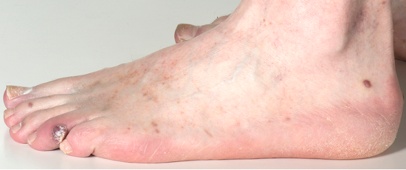
A 63-year-old Caucasian male was observed to have a lesion with overgrown tissue on the left 4th toe during a routine foot examination in the general diabetes clinic. He was given a short course of antibiotics and referred to the foot clinic for further evaluation. His medical background included type 2 diabetes mellitus, which was being managed with insulin therapy, hypertension, and a raised body mass index (BMI). His grandparents were from Malta indicating Mediterranean ancestry. On examination, he presented with a raised violaceous lesion on the lateral aspect of the proximal interphalangeal joint of the fourth toe. Sensations were adequate to 10gm monofilament and pulses were palpable. An X-ray of the foot was unremarkable. Due to unusual nature of the wound, a systemic examination was performed, which revealed purplish plaques on the forearm. Left foot radiograph in lateral and dorsoplantar views showed normal soft tissue and bony alignment with no osteolytic lesions of the fourth digit.
Figure 1 shows a raised lesion with a purplish hue on the lateral aspect of the left fourth toe which was found during a routine foot examination in the general diabetes clinic.
Investigations
As the presentation did not fit the classical DFU pattern, a referral was made to dermatology for further assessment. He had a skin biopsy of a similar lesion on his forearm. Histological examination revealed, spindle cells on immunohistochemistry positive for CD31 and Kaposi’s sarcoma-associated virus (KHSV also known as Human Herpes virus 8 – HHV8). Due to this virus’ strong association to HIV-associated immunodeficiency HIV testing would be strongly indicated. After initial consideration, our patient eventually agreed to be tested for HIV. Immunoassays for p24 antigen and IgM and IgG antibodies to HIV-1 and HIV-2 viruses were negative. Repeat serology was also negative. A whole-body CT scan did not show any internal organ involvement.
Treatment and follow-up
He initially underwent a period of observation and treatment with topical alitretinoin 0.1% gel by dermatology but was subsequently referred to the regional oncology tertiary care centre for consideration of localised radiotherapy due to being refractory to treatment. In the initial months, he was kept under observation as some lesions on the left forearm regressed spontaneously. However, he subsequently developed multiple new lesions that warranted intervention. He had radiotherapy at 8gy in 2 fractions, 6MeV electrons and is planned for further courses pending development of any new or refractory lesions. It is important to note that Kaposi’s sarcoma is an incurable infection. Treatment is geared towards controlling symptoms, as well as preventing bleeding or metastasis to other areas of the skin and organs. Tight glycaemic control to decrease tissue vulnerability to Kaposi’s sarcoma is paramount for long-term remission and prevention of any associated complications.
Discussion
Differentiating diabetes related foot ulcers from other lesions
According to 2019 data, the global diabetes prevalence is 9.3% which equates to 463 million people worldwide. The lifetime risk of developing foot ulcers in a patient with diabetes is between 15–20% (Saeedi et al, 2019). In contrast, the incidence of Kaposi’s sarcoma in a HIV negative population is 1.53 per 100,000 person years (Liu et al, 2018). Hence, when Kaposi’s sarcoma occurs in patients with diabetes and manifests as a pedal lesion, it can sometimes be misdiagnosed as a DFU. A high index of suspicion is needed to get early histological confirmation in all non-classical presentations. The acronym ‘CUBED’ (coloured lesion, uncertain diagnosis, bleeding, enlargement despite standard treatment, delay in healing for more than two months), which was originally drafted to detect pedal melanoma, can be a useful tool in other suspicious lesions such as Kaposi’s sarcoma (Bristow et al, 2010).
In our case, due to the atypical presentation, unusual site and history of bleeding on minimal trauma, a systemic examination was carried out to look out for lesions elsewhere on the body. We noticed two purplish plaques on his forearm and hence referred him promptly to the dermatology team following his first visit to the foot clinic. Prompt histopathological evaluation allows for a definitive diagnosis as other lesions such as DFUs can mimic the appearance of Kaposi’s sarcoma lesions due to its polymorphic variability. Other mimics could include melanoma, fungal infections and other infectious skin lesions such as mycetoma or bacillary angiomatosis for which diabetes is a risk factor.
Possible links between diabetes and Kaposi’s sarcoma
Kaposi’s sarcoma is a malignant condition of the endothelial cells of the blood vessels and the lymphatic system that is associated with Kaposi’s sarcoma-associated virus (Mesri et al, 2010). Classic Kaposi’s sarcoma mainly occurs in elderly men of Mediterranean ancestry, as seen in our patient. It is one of the most common cancers in HIV-infected individuals but its association with diabetes mellitus is not clearly understood in the HIV-negative cohort. Several acute inflammatory diseases are associated with Kaposi’s sarcoma-associated virus. Once infected with the virus, the host immune system will try to eliminate the infection by recognising the pathogen and developing an adaptive response (Mesri et al, 2010). However, the virus tends to escape the host defence by expressing an array of proteins and small RNA, which evades an immune response and the host eventually establishes a lifelong latent infection (Purushothaman et al, 2016). Kaposi’s sarcoma-associated virus will then work its way to dampen the immune system further to begin in the lytic cycle from which the virus can proliferate. A suitable environment such as an immunodeficiency state from AIDS helps the virus to express its genes that ultimately results in tumorigenesis (Purushothaman et al, 2016). There is growing evidence to substantiate the reactivation of the latent Kaposi’s sarcoma-associated virus in the hyperglycaemic environment. Impaired microcirculation in patients with diabetes results in tissue hypoxia. This may trigger the Kaposi’s sarcoma-associated virus lytic cycle through hypoxia-inducible factor alpha (HIF-alpha). HIF alpha can also be stimulated by IGF-1 factor (Davis et al, 2001; Catrina et al, 2006). Streptozocin injected hyperglycaemic mice when infected with Kaposi’s sarcoma-associated virus showed a high level of viral lytic gene expression compared with normal mice. This then led to Kaposi’s sarcoma-like tumours. It is believed the high H2O2 produced in the hyperglycaemic environment activates the mitogen activated protein kinase pathways (MAPK) that ultimately reactivates the latent Kaposi’s sarcoma- associated virus. The involvement of histone hyperacetylation of viral chromatins has also been suggested (Ye et al, 2016; Chang et al, 2017).
Stigma attached to HIV testing and delay in diagnosis
Despite attempts by our diabetes and dermatology team, our patient had considerable deliberations regarding HIV testing. He felt that the test unnecessarily challenged his marital fidelity, as he had always been faithful to his wife. After a detailed explanation about the difference in treatment between positive and negative tests, he reluctantly agreed to have the test, which turned out to be negative. Stigma related to AIDS is prevalent and contributes to low testing rates. In the UK, around 6% of the population living with HIV are undiagnosed (Chadwick et al, 2022). The Current UK General medical council does not allow implied consent as an option for HIV testing. Although this was not the case in our patient, delay in diagnosis often results in avoidable mortality and morbidity (Celesia et al, 2013).
Radiotherapy
Various factors, including the extent of the disease and the comorbidities determine the management approach. In general, all forms of Kaposi’s sarcoma show a decent response to radiotherapy ranging from 47% to 99% when a dose between 20Gy and 30Gy is used. Therapeutic indications for radiotherapy use in Kaposi’s sarcoma are detailed in National Comprehensive cancer network (NCCN) guidelines (Reid et al, 2019; Quéro et al, 2022). Our patient began radiotherapy at 8gy in 2 fractions, 6MeV electrons and is planned for further courses due to refractory disease.
Conclusion
In summary, we have presented a case of a pedal manifestation of Kaposi’s sarcoma that was initially thought to be diabetes-related foot lesion. Although Kaposi’s sarcoma is usually considered to have pathognomonic presentation and clinical features, when such lesions manifest in the foot, especially in patients with diabetes, it is not uncommon for the diagnosis to be mistaken for diabetes-related foot ulcers. This case highlights the importance of carrying out a general physical examination to identify possible systemic features and early histological confirmation of all atypical diabetes-related foot wounds.