This meeting report is based on a ‘Made Easy’ workshop that took place at the Wounds UK Annual Conference in Harrogate, on 12th November 2024. The workshop and meeting report were sponsored by an educational grant from Medicareplus International.
The first part of the workshop was presented by Sharyn Harte, and included the following key areas:
- The pathophysiology and different types of moisture-associated skin damage (MASD).
- The complexities of MASD and the importance of improving patient outcomes.
In the second part, Ayesha Marshall presented her latest publication, Stoma Care: Made Easy, as a go-to resource for clinicians caring for skin complications in people living with stomas. Highlighting practical tips to identify these complications, she shared easy-to-apply solutions for early identification and treatment.
In the UK, there is a significant unmet need for easy-to-access and practical solutions for clinicians in assessing and classifying MASD. A structured approach is needed to devise a prevention and treatment plan, tailored to each patient’s needs. This practical session helped provide guidance on this structured approach to the attending clinicians for understanding, diagnosing and managing different types of MASD.
The pathophysiology of MASD
The skin is the largest organ of the body and serves several crucial physiological functions. It maintains homeostasis, acts as a chemical and physical barrier to protect the body and provides immunity (Moncrieff et al, 2015). If skin integrity is compromised, the loss of these crucial functions can result in skin damage, wounds and, potentially, infections, causing a significant loss of patients’ quality of life (QoL) due to pain, anxiety, reduced mobility, malodour and/or other morbidities (Moncrieff et al, 2015).
With ageing, the barrier functions of the skin become impaired and the skin becomes vulnerable (Murphree, 2017). Figure 1 depicts the main layers of skin which play a crucial role in its functions. Patients with compromised skin integrity, therefore, are at increased risk of developing skin damage. MASD is a common form of skin damage in vulnerable patients, which occurs when the skin is exposed to excessive moisture for long periods of time. This disturbs the acid mantle function and results in loss of skin integrity, particularly in patients where the skin is already vulnerable (Parnham et al, 2020). As a result, irritants can penetrate the epidermis and the skin becomes prone to damage, such as tears and inflammation.
The pH of skin and the acid mantle
The outer layer of the epidermis (called the upper stratum corneum) contains a buffer system that maintains the skin’s acidic pH (Brooks et al, 2025). This buffer system is called the ‘acid mantle’, and it maintains the normal skin pH of 4.5–5.75. Created by a mixture of secretions from the sebaceous glands on the skin’s surface, it has important functions in keeping normal skin permeability, controlling inflammation and providing a defence against microbial attacks. A dysfunctional acid mantle results in dermatological disorders, making it an important therapeutic target in skin disorders (Brooks et al, 2025).
Types and diagnosis of MASD
MASD can be broadly divided into four categories:
- Incontinence-associated dermatitis (IAD).
- Periwound moisture-associated dermatitis.
- Intertriginous dermatitis (ITD; also known as intertrigo).
- Peristomal moisture-associated dermatitis.
Incontinence-associated dermatitis
IAD results from prolonged exposure to moisture from urine and faeces, both of which increase the pH of the skin (National Institute for Health and Care Excellence [NICE], 2024). It is generally hard-to-treat and can cause significant distress and pain for patients. Although it is not an uncommon condition, IAD can be under-reported or inappropriately diagnosed. Up to 1% of all adults may face regular incontinence (NICE, 2024), and up to 14 million people in the UK (including adults and children of all ages) have some form of urinary incontinence (NHS England, 2018).
Despite this, there are no centrally collected UK data on the prevalence of IAD or the number of patients who require continence-related care. Therefore, it is crucial that frontline clinicians and support staff understand prevention, identification and treatment of IAD. A key aim of this Made Easy session was to provide practical knowledge about diagnosing IAD, and educate the participating clinicians on optimal prevention and treatment techniques (see Figure 3, page 45, for further practical information).
By definition, IAD occurs in the genital areas (between skin folds/crevices; Banharak et al, 2021). Skin discolouration is typically the first indication of developing IAD. It is important to remember that discolouration may present differently in people with different skin tones (Dhoonmoon et al, 2023). Sharyn Harte presented a skin colour assessment tool [Figure 2] that can be used to assess the ‘normal’ (baseline) skin tone in people with suspected IAD.
IAD can cause significant, burning pain, especially as the patient experiences each episode of incontinence. This likely happens due to the exposure of the dermal-epidermal nerve endings to the irritants [Figure 1]. The exposure happens due to breaks in the skin after persistent moisture exposure. Clinicians and support staff can use some basic questions to investigate if there is suspicion of IAD:
- Does the affected area feel different to the surrounding skin?
- Can you see widespread, diffuse and blotchy erythema?
- Are there any patches of skin denudement or partial-thickness erosions?
- Is the patient experiencing pain, itching, change in sensation?
- Is the area warm to the touch?
- Is the affected area swollen?
- Do you notice any leakage of serous exudate or signs of bleeding?
It is important to remember that erythema can appear as redness in less deeply pigmented skin. However, in people with black, brown and olive skin tones, darkening/discoloration may be more subtle. Erythema can also appear as pink, red or purple areas in the skin that is exposed to irritants.
It is also important to remember that the damage may be in skin folds or over a bony prominence, natal cleft or as a peri-anal irritation with irregular-shaped edges.
Sharyn Harte explained that there is still significant confusion among clinicians about the differences between IAD and pressure ulcers (PUs), potentially leading to misdiagnosis/late diagnosis. IAD can make the skin vulnerable to PUs and, if not diagnosed correctly and in a timely manner, can be mistaken for PUs. However, these two conditions can also co-exist. Figure 3 depicts the major differences between PUs and IAD. It is crucial to note that, if the damaged skin contains necrotic tissue, it is highly likely that the patient has both a PU and IAD. This patient’s case should be recorded and treated as a PU.
Intertriginous dermatitis (ITD)
ITD occurs due to entrapment of sweat in skin folds that have limited or no air circulation (Kalra et al, 2014). This trapped sweat and associated irritants trigger a localised inflammatory reaction, known as ITD (or ‘intertrigo’). To diagnose ITD, it is important to remember that it is the trapped sweat that makes the local skin vulnerable. Overhydration and maceration in skin folds results in friction between the folds, so the damage is often mirrored on both sides of the fold — an identifying characteristic depicted in Figure 4A.
Periwound moisture-associated skin damage
Wounds produce exudate as part of the normal healing process, resulting from the inflammatory stage of wound healing. However, excessive exudate causes overhydration of the surrounding skin. This impairs the acid mantle and results in maceration and/or excoriation (Holloway, 2019). Depicted in Figure 4B, this damage occurs mostly due to the corrosive effects of proteolytic enzymes in the exudate. Compared to acute wounds, the exudate from non-healing wounds can cause more skin damage and contain more corrosive elements. To treat this type of skin damage, it is important that the fluid retention capacity of the wound dressing is appropriate for the exudate level so that excessive moisture can be controlled.
Peristomal moisture-associated dermatitis
Ayesha Marshall presented this section and introduced her latest publication, entitled ‘Stoma Care: Made Easy’ (Wounds UK, 2024).
By definition, a stoma is “an artificial opening made into a hollow organ, especially one on the surface of the body leading to the gut or trachea” (Wounds UK, 2024). Peristomal moisture-associated dermatitis occurs when the skin around this stoma is exposed to moisture leakage from the stoma. With 21,000 new cases reported each year in the UK alone, stoma care is a major burden for both clinicians and patients (Rolls et al, 2023).
For the purpose of this Made Easy workshop, the focus of Ayesha’s talk was on stomas that are created in the abdomen to remove urine or faeces from a patient’s body when the bladder or bowel cannot perform their excretion function (Wounds UK, 2024). In people living with these abdominal stomas, any leakage from the opening exposes the surrounding skin to urine or faeces, which leads to loss of skin function over time. This is called peristomal moisture-associated skin damage (PMASD), a chemical injury that compromises the function of stoma itself, if not treated in time (Burch et al, 2021). In addition, people with a stoma routinely need to change their stoma pouches. If appropriate care is not taken in this removal process, or if the skin around the stoma is fragile due to irritant exposure, the removal of the pouch can damage the skin. This is called peristomal medical adhesive-related skin injury (PMARSI) and is often under-reported (LeBlanc et al, 2019).
Together, these two stoma-related skin injuries can cause significant distress to the patient and are costly to treat. In the UK, each severe case of peristomal complication can cost over £500 to treat and every person living with a stoma faces at least one episode of a peristomal complication over their lifetime (Rolls et al, 2023). Therefore, there is an unmet need for timely identification and treatment of these issues.
Having highlighted these significant challenges, Ayesha Marshall shared key identification and diagnosis tips that can help clinicians identify PMASD and PMARSI in a timely manner [Table 1].
Treatment of different types of MASD — a unifying step up/down approach
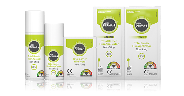
Sharyn Harte shared a total barrier protection (TBP) treatment strategy, supported by two case studies, to illustrate the effectiveness of the Medi Derma-S and Medi Derma-PRO product range in effectively managing MASD. Figure 5 shows how this step up/down approach can be employed, tailored to the severity of MASD.
The Made Easy session ended with a knowledge-check quiz to assess how the participants could apply their learning from the session to differentiate between cases of PUs versus IAD. Over 95% of all attending clinicians were able to successfully differentiate the two conditions for each image shown on the screen, demonstrating a successful learning outcome.
Conclusion
Skin breakdown associated with MASD can cause huge distress and loss of QoL to patients, and is a significant burden for clinicians and healthcare systems. Furthermore, MASD management requires long-term prevention and treatment approaches. In the UK, MASD has a high incidence in the ageing population, in care home facilities and in hospitalised patients (NHS, 2018).
This Made Easy session raised awareness of peristomal MASD and the unmet need in timely identification of different types of MASD, providing practical tips for both diagnosis and treatment. MASD tends to be under-reported, potentially leading to higher treatment costs downstream.
The Medicareplus Medi Derma-S and Medi Derma-PRO range used as Total Barrier Protection™ treatment strategy can help clinicians improve this situation by providing options suitable to each level of MASD severity. With specific and timely interventions, it is possible to reduce MASD incidence in the UK care setting.