This meeting report is based on a ‘Made Easy’ workshop that took place at the Wounds UK Annual Conference in Harrogate, on 12 November 2024. The workshop and meeting report were sponsored by an educational grant from CliniMed Ltd.
The workshop was opened by Dr Jeannie Donnelly (UK Director ISTAP; Lead Nurse, Senior Honorary Lecture) who outlined the learning objectives of the session:
- Raising awareness of skin tears and the importance of identifying at-risk patients.
- Exploring strategies for prevention and risk reduction.
- Understanding the causes of a medical adhesive-related skin injury (MARSI).
- Highlighting reasons for using sterile medical adhesive to reduce the risk of skin tears and injuries.
What are skin tears?
There have been several definitions and classification tools proposed for skin tears. The most well-researched definition is the International Skin Tear Advisory Panel (ISTAP), which should be used0 for the identification and classification of a skin tear. This states that a skin tear is: “a traumatic wound caused by mechanical forces, including removal of adhesives. Severity may vary by depth (not extending through the subcutaneous layer)” (LeBlanc et al, 2018).
Skin tears occur in individuals with fragile skin, commonly neonates and older people. Studies have shown that the prevalence of skin tears is higher than that of pressure ulcers (Carville et al, 2007), but compared to pressure ulcers, skin tears are often underestimated and considered largely preventable. They can occur on any part of the body but most (50-80%) occur in the upper extremities (arms and hands), followed by the lower extremities (15-57 %), with 5% occurring in other areas of the body (Carville et al, 2014, Sanada et al, 2015; LeBlanc et al, 2011, 2020).
Patients suffering from skin tears report increased pain and distress, with decreased quality of life (LeBlanc et al, 2018). In addition, these patients are also at an increased risk of developing infections and comorbidities, which may lead to increased healthcare costs (Rayner et al, 2015) and contribute to longer hospital stays (Carville et al, 2014; LeBlanc et al, 2016).
Causes of skin tears
The causes of skin tears can vary, with almost 50% of cases occurring without any apparent cause. However, common factors include:
- Blunt trauma.
- Activities of daily living.
- Dressing or treatment-related injuries.
- Falls.
- Injuries during patient transfer (caused by friction, shear and poor handling).
- Equipment-related injuries (e.g., wheelchairs, side rails, and beds).
Risk factors for skin tears
Dr Jeannie Donnelly used the analogy of a plane breaking through the sound barrier to explain the concept of breaking the skin barrier. She introduced the acronym SOUND to describe key factors affecting the skin barrier:
- S: Skin age.
- O: Oxygen/perfusion.
- U: Underlying factors.
- N: Nutrition and hydration.
- D: Diseases and drugs.
Skin age
Neonates, particularly premature infants with a very low birth weight (less than 1,500 g) or extremely low birth weight (less than 1,000 g), have fragile, underdeveloped skin that is easily traumatised. At full term, the skin is only 60% of adult thickness, and neonatal skin is less elastic, making it more susceptible to damage from shear forces (Irving et al, 2006). This underdeveloped skin barrier leads to high water loss, increased permeability to external agents, and increased risk of infection.
Neonates and premature infants often require intensive medical care and support, which, while essential for their survival, can inadvertently damage their skin. For example, the use of adhesives in dressings, electrode tape, or device securement – such as adhesive tapes for nasogastric (NG) tubes, combined with oxygen therapy and other monitoring equipment, may increase the risk of injury to their already vulnerable skin.
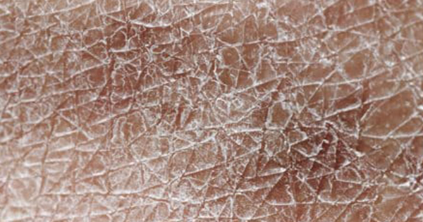
On the other end of the spectrum, older adults are more prone to skin barrier breakdown due to the natural ageing process. Ageing skin is more fragile and susceptible to damage, requiring less force to cause traumatic injury compared to individuals with healthy (Voegeli, 2007). This increased vulnerability is associated with age-related changes in the skin, such as atrophy, ecchymosis, senile purpura, haematomas, stellate pseudoscars, and photodamage from sun exposure. Dry skin (xerosis) and advanced age often result in itchy skin (pruritus), which can lead to mechanical trauma caused by scratching. Additionally, ageing (and associated co-morbidities) reduces skin moisture, decreases sensation and delays wound healing, further increasing the risk of skin injuries (Moncrieff et al, 2015).
Oxygen/perfusion
Inadequate oxygenation and impaired tissue perfusion can weaken the skin’s integrity, making it more susceptible to injuries like skin tears.
Underlying/lifestyle factors
Major pollutants affecting the skin (solar ultraviolet radiation, polycyclic aromatic hydrocarbons, volatile organic compounds, nitrogen oxides, particulate matter, cigarette smoke, heavy metals and arsenic), can act alone, synergistically or in combination, potentially leading to additive effects. These pollutants alter barrier function, elasticity, thickness, dermal extracellular integrity and skin absorption (Fitoussi et al, 2022).
Nutrition and hydration
Optimising nutrition and hydration is as important as minimising unintentional harm. This can be achieved by involving a dietitian for nutritional support, particularly for patients with extreme BMIs (<20 or >30), as are regular medication reviews, in collaboration with pharmacists or the medical team to reduce or optimise medications that may contribute to skin fragility.
Disease and medication
Conditions such as hypotension, inflammation, diabetes and immunosuppression, as well as the use of steroids and other medications, are known to affect skin health. Allergies and other medical conditions can also play a role, necessitating careful evaluation and management.
Additional factors that increase the risk of skin injuries include: individuals needing assistance with activities of daily living (e.g. bathing, dressing and mobility; Wounds UK, 2015); patients with a history of skin tears, which may leave characteristic crescent-shaped scarring; those who are acutely unwell, confused, or agitated; and patients with dermatological conditions (e.g. eczema, dermatitis, chronic exudative ulcers and epidermolysis bullosa).
Managing underlying factors
In patients with vulnerable skin, risk factors and incidence of skin tears are often associated with other underlying conditions. For example, consider a patient who develops a skin tear after stumbling and falling. Upon investigation, it may be found that the fall occurred due to dizziness. Managing the underlying cause of the dizziness – whether it is an ear infection, vertigo, dehydration, dementia, low blood sugar, stress, iron deficiency anaemia, migraine, motion sickness, postural hypotension, or a medication side effect – could significantly reduce the likelihood of future falls. Therefore, by addressing the root cause, healthcare professionals can help minimise the risk of skin tears. This approach also emphasises the importance of viewing skin tears not as isolated events but as part of a broader clinical picture that requires comprehensive assessment and intervention.
Classification of skin tears
Although no universally accepted classification system exists for skin tears, several tools are available. To address the need for a user-friendly and simple tool, ISTAP developed the systematic, standardised and validated ISTAP Skin Tear Classification System (LeBlanc et al, 2013), which has been psychometrically tested and classifies skin tears into three categories:
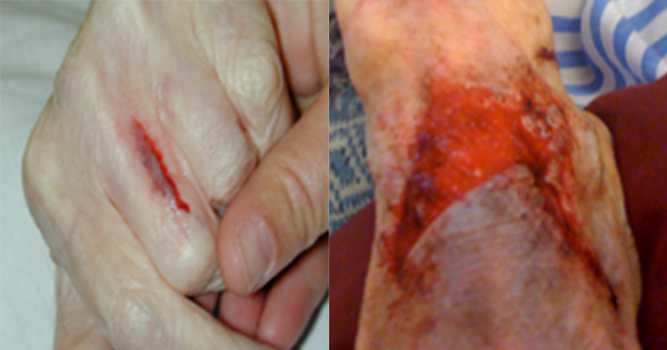
- Type 1 (no skin loss): Linear or flap tear which can be repositioned to cover wound bed [Figure 1].
- Type 2 (partial flap loss): Which cannot be repositioned to cover wound bed [Figure 2.
- Type 3 (total flap loss): Exposing the entire wound bed [Figure 3].
The validated classification system has been translated into numerous languages, and Van Tiggelen et al (2020) recommend that the ISTAP classification system be adopted into clinical practice as a universal language for describing and documenting skin tears.
Goals of treatment
Treating skin tears should aim to preserve the skin flap and maintain the surrounding tissue, reapproximate the edges of the wound (without stretching the skin), and reduce the risk of infection and further injury. The skin tear decision algorithm [Figure 4] is designed to help practitioners in the assessment and treatment of skin tears, maintaining a continuous link between preventing, assessing and treating skin tears.
Control bleeding and assess
Apply pressure and elevate the limb if possible. If the skin flap [Box 1] is present but not viable, it may need to be debrided. Care should be taken during debridement to ensure that viable skin flaps are left intact and fragile skin is protected.
Cleanse/irrigate the wound
Cleanse/irrigate the wound as per local protocol and remove any residual debris or haematoma [Figure 5]. Pat the surrounding skin dry gently to avoid further injury.
Reapproximate skin flap
Where possible, the edges of the skin flap must be brought together [Figure 6] to use as a ‘dressing’. Ease the flap back into place using a gloved finger, dampened cotton tip, tweezers or a silicone strip.
Dressing application
Once the flap has been approximated (if present), it should be secured with a dressing that promotes moist wound healing. The selected dressing should also be atraumatic on removal and help reduce pain.
Dressing removal
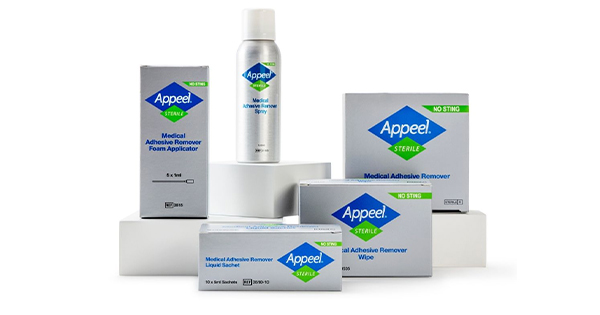
Skin tears may be associated with causing patients unnecessary harm, e.g. if the skin tear is sustained due to medical adhesive-related skin injury (LeBlanc et al, 2018). Therefore, during the dressing removal process, it may be necessary to use a sterile medical adhesive remover (e.g. Appeel Sterile; CliniMed Ltd.) to aid in removing a dressing.
It’s important to be aware of products that shouldn’t be used when you have a patient with a skin tear. See Box 2 for more information.
Focus on compression
When managing skin tears of the lower limb, it is recommended that compression should be considered as a component of treatment (LeBlanc et al, 2014, 2018; Ewart, 2016). The application of compression as an adjunct to wound therapy has been found to control peripheral oedema and address local swelling, which is recognised as a factor in delayed healing (LeBlanc et al, 2014).
If red flag symptoms/conditions [Box 3], immediately escalate to the relevant clinical specialist (National Wound Care Strategy Programme, 2023)
In the absence of red flags, it is recommended to start mild compression (<21 mmHg) in all patients with lower limb skin tears. This can be increased to strong compression (up to 40 mmHg) in patients who have had a full vascular assessment.
Inflammation versus infection: What is the difference?
Wound inflammation from trauma should be distinguished from wound infection. Inflammation is a normal part of the healing process in a wound. It manifests as typically swelling, heat and pain, and can occur even if the wound is not infected.
Infection occurs when pathogenic microorganisms, such as bacteria, enter the body and begin to multiply. Clinical signs of infection (e.g. swelling, heat and pain) overlap with those of inflammation. However, while inflammation will almost always occur when infection is present, it is possible to have inflammation without infection. Jeannie Donnelly emphasised the importance of being able to recognise and differentiate between inflammation and local infection, as this distinction is important in avoiding the overuse of antibiotics or antimicrobial topical treatments.
Diagnosis of infection should be based on clinical assessment including sending discharging fluid for gram staining, culture and sensitivity testing (a culture swab of a wound should only be taken if clinical infection is suspected; it is important to differentiate between the wound being infected versus contaminated).
Preventative care
Dr Jeannie Donnelly mentioned the importance of approaches to preventative care, which can significantly reduce the risk of skin tears, particularly when tailored to a patient’s individual needs and abilities.
General health
For patients with intact cognitive function, a self-management approach is often beneficial. This includes educating patients and carers on skin tear prevention, risks of skin fragility due to age or medication, medication-induced skin fragility, safe moving and handling practices, as well as encouraging their active involvement in treatment decisions, if appropriate.
Mobility
Encouraging active involvement in mobility, where possible, is an important element of prevention. The appropriate selection and use of assistive devices, along with physiotherapy or occupational therapy referrals, can improve mobility and support safe transfers. Proper manual handling techniques, such as using sliding sheets rather than, for example pulling patients up the bed, can prevent skin trauma during repositioning.
Falls are a major cause of skin tears; therefore, initiating a fall assessment and prevention programme is important. Simple measures, such as decluttering the environment around the patient’s bed, ensuring adequate lighting, and padding equipment like bed rails or wheelchairs, can further reduce risks. Proper footwear should also be assessed to promote safe movement.
Skin
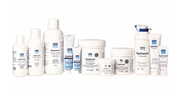
Maintaining skin integrity is at the core of preventing skin tears. Education patients and providing awareness of medication-induced skin fragility, especially with the use of topical and systemic steroids, is fundamental. Protective clothing or devices, such as long sleeves or shin guards, can be worn to safeguard and protect vulnerable areas. Regular skin moisturising, ideally twice daily, and maintaining short fingernails for both patients and carers can reduce trauma risks.
Good skin hygiene should include the use of warm or tepid water, avoiding soap that may dry the skin, and moisturising immediately afterward. Controlling conditions such as oedema, through measures like leg elevation, can also help maintain skin health. When dressings are required, avoiding strong adhesives and using a sterile medical adhesive remover during removal can minimise damage to fragile skin.
Medical adhesive-related skin injury
Stephanie Stanley (Consultant Podiatrist, Hampshire and Isle of Wight Healthcare, NHS Foundation Trust), went on to cover the objectives of understanding the causes of a medical adhesive-related skin injury (MARSI) and highlighting reasons for using sterile medical adhesive to reduce the risk of skin tears and injuries.
The most common cause of a MARSI is the removal of a dressing or device that contains a medical adhesive (mechanical forces). This type of injury is commonly encountered in podiatry due to the frequent application and removal of padding [Figure 7], dressings, and adhesive tapes, often required in managing wounds or other conditions (Fumarola et al, 2020).
Causes of MARSI
Continuous use of adhesive tape, particularly in patients managing chronic foot ulcers, can lead to skin irritation and damage during the removal process. This can result in [Figure 8]:
- Skin stripping.
- Skin tear.
- Tension blister.
- Contact dermatitis.
- Allergic dermatitis.
- Folliculitis.
Diabetic foot wounds and MARSI
Patients with diabetes are particularly vulnerable to MARSI due to the prevalence of diabetic foot wounds, which often necessitate frequent dressing changes and adhesive use. These wounds can be categorised into:
Neuropathic wounds: Typically located in load-bearing areas of the foot, these wounds result from pressure and a lack of sensation caused by neuropathy [Figure 9].
Ischaemic wounds: Often found on the periphery of the foot, these wounds result from poor blood supply. In such cases, clinicians should review ankle–brachial pressure index (ABPI) or toe brachial index (TBI) to assess vascular health [Figure 10].
Neuro-ischaemic wounds: These result from a combination of neuropathy and ischaemia, often presenting as complex wounds requiring multidisciplinary care [Figure 11].
Tips for dressing application and removal
Care needs to be taken when managing a lower limb skin tear to negate or minimise the risk of trauma to the wound or surrounding skin when applying and removing the dressing (LeBlanc et al, 2018). Particular consideration needs to be given to flap preservation: so, when applying the dressing, a good tip is to draw an arrow on the dressing to indicate the correct direction of removal [Figure 12]. Always remove the dressing in the direction of the arrow and the direction of hair growth.
Time should be taken to remove dressings slowly and carefully. Although there are dressings that are specifically designed to be atraumatic (e.g. lipido-colloid dressings), it may be necessary to use a sterile medical adhesive remover when removing the dressing to minimise trauma (LeBlanc et al, 2018).
Appeel Sterile Medical Adhesive Remover
Best practice guidance recommends the use of sterile silicone medical adhesive removers, such as Appeel Sterile.
When Appeel Sterile comes into contact with the adhesive of the dressing, it temporarily changes the surface energy of the skin, disrupting the adhesive properties between the skin and device and loosening the bond without compromising the surrounding area. Features of Appeel Sterile include:
- Sterile – designed for use on both intact and injured skin; does not impair wound healing
- Solution for all wound types: Extensive range of products
- Easy to use
- No-sting formula
- Fast drying and leaves no residue.
It aids in the removal of:
- Adhesive dressings
- Adhesives securing devices such as central lines, nasogastric tubes, intravenous and subcutaneous cannulae.
For good clinical practice, please refer to the indications for use for Apppeel sterile.
Top tips for the application of Appeel Sterile Medical Adhesive Remover
With the unique formula and only sterile adhesive remover on the market, Dr Jeannie Donnelly and Stephanie Stanley gave the audience some practical tips that they have learnt in their practice, as seen on the next page.