Journal Articles

Photo mapping foot deformity: a picture speaks a thousand words

Share this article

Photo mapping foot deformity: a picture speaks a thousand words

Abigail Platts
6 November 2020

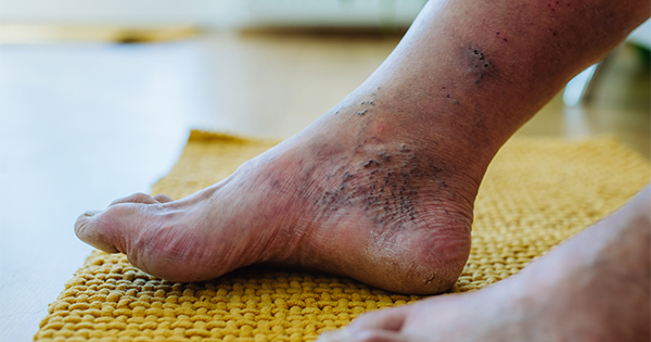
The use of photography in podiatry is well established within wound care practice, the benefits of which are also widely documented. In recognising the role that photography plays in documenting ulceration, it became clear that the benefits need not be limited to this area of podiatry practice and that its use would be a valuable tool in the mapping of deformity of the foot. The use of photography has therefore been expanded into this area. The aim of these photographs is to support the written documentation within the Musculoskeletal (MSK)/High Risk assessments, including non-weight-bearing and weight-bearing assessment of the feet. Mapping deformity is key to being able to accurately monitor foot changes and potential deformity. Although patients’ notes have a written description of the foot, the use of supporting photographs help to map small and important changes over time and give an immediate visual comparison.

Free for all healthcare professionals

Sign up to the Wounds Group journals


By clicking ‘Subscribe’, you are agreeing that the Wounds Group are able to email you periodic newsletters. You may unsubscribe from these at any time. Your info is safe with us and we will never sell or trade your details. For information please review our privacy policy.

Are you a healthcare professional? This website is for healthcare professionals only. To continue, please confirm that you are a healthcare professional below.

We use cookies responsibly to ensure that we give you the best experience on our website. If you continue without changing your browser settings, we’ll assume that you are happy to receive all cookies on this website. Read about how we use cookies.

I am not a healthcare professional.