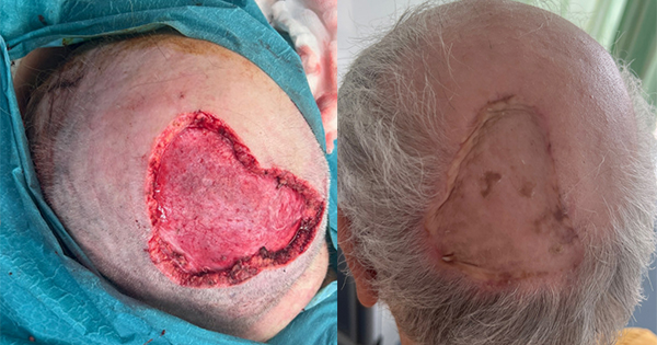
Scalp and forehead reconstruction constitutes a significant clinical challenge, given the intricate complexities inherent in the disease process, the distinct properties of the tissue designated for reconstruction, and the elevated postoperative expectations, especially considering the prominent positioning of the forehead and scalp in facial aesthetics. Oncologic resections, secondary to skin cancers and intracranial lesions, predominantly account for scalp defects that require reconstruction. Nonetheless, a myriad of other etiological factors also contribute to the prevalence of such defects, encompassing trauma, burns, infections, radio necrosis and congenital anomalies (Shonka et al, 2011).
Reconstruction follows a stepwise approach through the reconstructive ladder [Figure 1] to address the size and depth of the defect. The approach is usually tailored with consideration of patient factors while minimising disturbances to surrounding tissue and maintaining both perfusion and innervation. In skin cancer surgery, achieving complete excision margins is the ultimate priority prior to reconstruction. Moreover, brow symmetry, contour and natural hairline should be maintained whenever possible to ensure satisfactory cosmetic outcomes are achieved (Angelos et al, 2009).
The scalp is a complex anatomical structure that comprises five layers recognised by the mnemonic SCALP: Skin, Connective tissue, Aponeurosis, Loose areolar connective tissue and Periosteum. The presence of this connective tissue layer (the galea), which is a rich dense vascular network with ample blood supply, lends to its unique healing process and supports different reconstructive planning. This vascular network is supplied by branches from both the internal carotid, through the supratrochlear and supraorbital arteries and external carotid arteries, through the superficial temporal, posterior auricular and occipital arteries, as it creates an extensive anastomotic arrangement within the subcutaneous layer.
The innervation of the scalp is as abundant as its vasculature and involves various nerves lending to its motor and sensory function. Motor input to the frontalis muscle is supplied by branches of the facial nerve, whereas sensory inputs are provided by the supratrochlear and supraorbital nerves, the zygomaticotemporal and auriculotemporal nerves and branches of dorsal rami of cervical spinal nerves and from the cervical plexus.
Important considerations should be taken while planning the reconstructive approach to avoid injuring these structures, positioning incisions at the borders of facial aesthetic units and avoiding traversing them.
General considerations
Conserving as much native scalp tissue as possible is paramount in scalp reconstruction. An equally important principle is respecting hair growth pattern and hairlines. Incisions should also be made parallel to the shaft of exiting scalp hairs to minimise hair follicle damage and consequent alopecia, and allow for later camouflage of scarring (Angelos et al, 2009).
The ideal replacement of scalp tissue is scalp tissue itself. An ideal aesthetic reconstruction, therefore, should involve the manipulation of residual scalp tissue as opposed to grafts or free flaps. The choice of the reconstructive technique is influenced by multiple factors as shown in Table 1 (Leedy et al, 2005) in an appropriate and timely manner to ensure patients were reviewed once the immediate care need was addressed.
Algorithm for scalp wound management
The surgical approach to scalp wounds should be tailor-made for each patient. Several algorithms [Figure 3] have been put forward for managing scalp defects depending on many factors. Leedy et al (2005) proposed an algorithm based on the size and location of scalp defects. However, a comprehensive algorithm should also address other patient factors such as comorbidities, age and scalp hair patterns. This was described by Cherubino et al (2013), who designed an algorithm
[Figure 3]that not only looked at the wound structure but also addressed comorbidities as well as different clinical situations [Table 2]. This should assist in making an informed choice, which ultimately remains highly individualised based on each patient’s specific needs.
Primary closure
This is usually possible for small defects less than 3cm. Good results are attained with tension-free closure. Excessive tension wound closure can cause alopecia from either hair follicle loss or anagenic phase arrest. Closure is done in layers or with a running interlocking suture taking the galea. The galea is responsible for most of the resistance to scalp flap advancement. Additional advancement can be achieved by carefully scoring the galea perpendicular to the direction of advancement. Each of these incisions results in approximately 1.67mm of tissue gain (Raposio et al, 1998). Care should be taken to prevent accidental injury to scalp arteries that lie just superficial to the galea.
Vacuum-assisted closure
Vacuum-assisted closure (VAC) for wound closure is widely used in surgery. VAC systems facilitate wound healing by increasing dermal and subdermal perfusion, decreasing oedema, lowering the bacterial load on wounds and stimulating granulation tissue growth. On the scalp, VAC is utilised in cases where granulation tissue growth encouragement is required before grafting and to bolster dressings after split-thickness skin grafting (STSG). As Andrews et al
(2006) have described, VAC can be used as a temporising dressing after debridement of a contaminated wound and prior to reconstruction with a skin graft.
Skin grafts
Both full-thickness and split-thickness grafts offer viable and reliable solutions for scalp reconstruction with no significant statistical difference in terms of graft adherence, rate of complications, or overall outcome between these two options. The choice between these can be made based on the surgeon’s preference and the specific needs of the patient (Hilton et al, 2019).
While reconstruction with skin grafts is considered a relatively straightforward procedure, it comes with several disadvantages. One limitation is that it tends to be cosmetically inferior compared to other options,such as local flaps. Skin grafts also lack hair-bearing properties, which can affect the overall aesthetic outcome. Additionally, they are more susceptible to local ulceration, leading to a higher risk of partial or complete graft loss.
Scalp reconstruction using skin grafts also has certain limitations, including cases involving large defects exceeding 20cm2, irradiated skin, or when the patient retains significant hair growth. In such situations, opting for local flaps would be a more suitable choice for achieving better outcomes.
The size of the defect will determine what type of skin graft could be used. Full-thickness skin grafts could be used in smaller defects, while split-thickness skin grafts could be a good option for defects between 10–20cm2 (Cherubino et al, 2013). Securing small full-thickness skin grafts with foam tie-over dressing was found to be associated with superior graft adherence and take compared to securing with sutures only (Sun et al, 2021), while NPWT can be used in larger defects reconstructed with STSG.
Full-thickness skin grafts can provide excellent cosmetic outcomes on the scalp due to reduced secondary contraction. However, their success depends on receiving abundant nourishment from the wound bed to achieve proper graft take. Therefore, the availability of an adequate wound bed is crucial for successful healing and graft integration.
Local flaps
Local flaps [Figure 5] are widely used in reconstructing forehead and scalp defects. They are advantageous in providing good colour, texture and depth match while minimising scars and maintaining hair alignment.
The most used patterns of local flaps in scalp reconstruction are transposition and rotational flaps with their various modifications. Advancement flaps are very limited in the scalp due to the poor elasticity and the multidirectional lines of tension of the underlying galea. Larger wide-based flaps are preferred over smaller multiple flaps due to increased reliability and fewer scars.
The decision-making in locoregional flap reconstruction depends mainly on two factors: the size and the location of the defect.
The vertex is the least elastic part of the scalp, additional considerations are required when choosing the appropriate locoregional flap options. The most widely used local flaps here are the triple limberg flap with its rotational variation, the pinwheel flap, and multiple O-to-Z flaps (Brawley and Sidle, 2022).
The triple Limberg flap
This is preferred by some authors as it can offer better distal flap tip vascularity with lesser risks of flap dehiscence. It is designed to close an approximate hexagonal defect. Three equidistant perpendicular lines are drawn from the defect’s edges, each is equal to one side of the hexagon. Three equidistant lines at approximately 60-degree angles are then drawn in a clockwise or counter-clockwise. After the incision and undermining, the flaps are rotated in the same direction to fill the defect.
The pinwheel flap
The pinwheel flap was first described by Vecchione and Griffith in 1978 and was proposed for small to moderate scalp defects. It is preferable as it offers minimal scarring and preservation of hair alignment while eliminating the need for a skin graft to cover the donor site.
Two variations of the pinwheel flap are described: two flaps (Ying yang variation) and three flaps (Isle of Man variation). The flap is designed to close a circular or oval-shaped pattern around the defect, with the central pivot points positioned in a healthy area with a good blood supply. The flap is incised along its limbs and the galea is scored conservatively along the tension lines to preserve the blood supply while providing elasticity to assist the flap inset (Varnalidis et al, 2019).
The O-to-Z flap
A double rotation flap commonly used for closing circular defects. The nature of this flap makes it a good option for reconstructing Vertex and temporoparietal defects in the hair-bearing area where hair can be used as a camouflage for the suture line. The flap design involves the rotation of two opposing curvilinear pedicles towards each other, effectively filling the defect and resulting in a suture line that resembles a Z shape.
The posterior cervical rotational flap
This flap is widely relied upon for reconstructing small to medium-sized occipital scalp defects. The flap design involves making a curvilinear incision, which can extend up to six times the diameter of the defect followed by deep undermining. This flap has proven to be highly successful, providing favourable tissue colour and texture matching, and is less cosmetically sensitive to patients.
The Orticochea flap
A versatile surgical technique that can also be adapted for reconstructing large defects in frontal or occipital scalp reconstruction. The Orticochea flap offers several benefits. The design allows for the closure of relatively large defects, providing ample tissue for advancement. It minimises tension on the wound edges hence, reducing the risk of wound dehiscence. This flap also offers a better cosmetic outcome as it provides a good colour and texture match with the surrounding scalp tissue (Mendoza et al, 2021).
While advancement flaps have limited applicability on the vertex and parietal scalp, they remain highly advantageous for cosmetically sensitive areas, such as the frontal scalp and forehead defects. Among these, the double opposing rectangular flap, known as the H-flap, is a commonly used technique for reconstructing small to medium-sized defects in this area. The horizontal incision lines of this flap can be camouflaged along the resting skin tension lines of the forehead, resulting in excellent cosmetic outcomes. Moreover, it offers optimal alignment for the eyebrow and frontal hairline.
Several other flap variations, including V-Y, bilobed, Limberg and rotational advancement, can also provide favourable results. However, extra attention and precise undermining before the flap inset are crucial to achieving proper alignment of the eyebrows and frontal hairlines for an aesthetically pleasing outcome.
Tissue expansion
Tissue expansion has a considerable role as a secondary procedure where local tissue rearrangements are inadequate to cover defects. Approximately 50% of the scalp can be reconstructed with expanded scalp tissue without changing the hair growth patterns or creating a new donor defect (Manders et al, 1984). When using expanders, it is generally recommended to use a single and the largest possible expander that fits to reduce the risk of infection associated with multiple expanders and procedures. However, the use of more than one expander is often necessary for scalp reconstruction.
The shape of the expander has a bearing on the amount of tissue gain. Round-based expanders give tissue gains of up to 25% while crescentic and rectangular-based ones may attain as much as 32% and 38% respectively (Van Rappard et al, 1988). The tissue defect should, however, dictate the type of expander to use. The expanded skin may be re-expanded to cover large defects without compromising the cosmetic quality of the scalp. To cater for tissue contraction during advancement, it is recommended to expand the skin by an extra 20% beyond the size of the defect.
This technique has the downside of requiring staged operations that have long interval periods, as well as a complication rate that ranges from 6–25% (Azzolini et al, 1992; Kuwahara et al, 2000).
Free flaps
Free tissue transfer provides durable, reliable cover in scalp reconstruction. The choice of
free flap reconstruction depends mainly on the size and location of the defect while also considering the patient’s general status. Currently, reconstructive flap choice relies on low-level evidence, expert opinions, and accumulated experience.
Free flaps are preserved for larger scalp defects of more than 30cm2 and those with exposed pericranium, dura or metal work as titanium mesh. The most common donor sites for these flaps are the anterolateral thigh (ALT), latissimus dorsi (LD), radial forearm (RFA), parascapular, and omental flaps There are advantages and disadvantages to each approach, as will be discussed below.
Anterolateral thigh (ALT) flap
The ALT flap[Figure 6] is designed along the outer border of the lateral thigh based on the blood supply from the lateral femoral circumflex artery. The design versatility of the ALT flap offers a significant advantage as it can be used as an adipocutaneous, fasciocutaneous, chimeric, or musculocutaneous flap to fit any reconstructive need. When combined with its accessibility in the supine position, having a longer pedicle, and minimal donor site morbidity, it leads to better patient outcomes and decreased operating times (Collins et al, 2012). The tensor fascia lata can also be used in conjunction as a free graft or an attached vascular graft which can also be used in dural reconstruction, confers an additional advantage.
Latissimus dorsi (LD) flap
As Gp M, Stueber K et al (1978) first described the use of the free LD flap for head and neck reconstruction, the flap can be raised either as muscle flap or with the overlying skin as a
myo-cutaneous flap. One of the main advantages of an LD flap is the potential size of the flap. When combined with its ability to further expand using tissue expanders, it can be used for subtotal or total scalp loss. It also can be very helpful in patients with small skull defects where it can be harvested with a rib portion that opens various avenues for its use within calvarial reconstruction (Xiao et al, 2020).
Parascapular flap (Figure 7)
First documented by Hamilton et al (1982), the parascapular flap has gained recognition as a reliable and adaptable choice for reconstructing large defects in the scalp and forehead. This flap consists of skin and subcutaneous tissue and is designed along the lateral border of the scapula, based on the blood supply from the vertical terminal branch of the circumflex scapular artery.
In a comparative study by Klinkenberg et al (2013), 60 patients who underwent free-flap reconstruction with either anterolateral thigh, para-scapular, or lateral arm flaps were included and followed for an average of 50 months. The study indicated greater patient satisfaction with the parascapular
flap compared to the anterolateral thigh
and lateral arm flaps.
The omental flap
First described as a free tissue transfer for scalp reconstruction by Mclean and Buncke (1972), the omental flap offers several benefits over other flap options. It is notably large, allowing for the reconstruction of substantial defects, even total defects and its thin, pliable nature makes it particularly well-suited for scalp repair. Its use is generally restricted due to concerns about donor-site morbidity and the potential for abdominal wall hernia as it is harvested through laparotomy incisions or endoscopic where possible. As a result, the omental flap is often reserved for instances where other flap options are either contraindicated or not readily accessible.
Flap comparison
In a study by Del Castillo et al (2021), several characteristics were identified as crucial for free flaps used in scalp reconstruction. These include adequate tissue volume, low donor site morbidity, a long vascular pedicle, reliable anatomical features, shape versatility and satisfactory aesthetic results. In this study, outcomes from scalp reconstructions using latissimus dorsi (LD), anterolateral thigh (ALT), and omental (OM) free flaps were compared.
The unique properties, along with the advantages and disadvantages of each available free flap, are detailed in [Table 3]. The optimal choice is highly individualised, depending on the specific needs of each patient. Intriguingly, while factors such as radiation exposure, prior surgical interventions and the presence of malignancy can adversely impact the success of free flaps, age does not appear to be a significant factor (Simunovic et al, 2016).
Conclusion
Scalp wound reconstruction is a formidable challenge and requires a systematic and comprehensive patient-centred approach that takes into consideration the size and location of the defects, the comorbidities, and the aesthetic requirements of each patient. A good understanding of the different reconstructive options when combined with respect to the anatomy and the aesthetic sub-unit, are the ultimate factors that contribute to both satisfactory functional and aesthetic outcomes.


