The treatment of peripheral arterial disease (PAD) and Chronic Venous Insufficiency (CVI) is not straightforward. Contradictory guidance on calculating the severity of arterial disease and safe provision of compression therapy (Adriennson et al, 2017), has led to uncertainty, where, through fear of doing harm, patients are referred for vascular assessment, rather than being treated (Elwell and Sneddon, 2019). Delays in preventative management lead to deterioration and leg ulceration (Atkin et al, 2021). These often require long-term wound management (Stanton, 2023), as 80% of ulcers recur within 3 months of healing, instigating a repetitive cycle (Probst et al, 2023); costing the NHS £8.3 billion per annum (Guest et al, 2020).
A mitigating factor in the inefficient treatment of recurrent leg ulcers is that they were once viewed as a venous pathology; more recently they were recognised as due to both venous and lymphatic insufficiency (Moffat et al, 2019). However, the picture is still incomplete, as the myofascial system and its role in wound healing has not been considered. Long-term healing can only be achieved by understanding and managing all inter-related body systems, which do not operate independently.
Lymphascial Kinesiology taping (LKT) is an application method for kinesiology tape (KT) that interacts with both myofascial and lymphatic systems (Anvar, 2016). It is an underestimated tool, which offers a low risk, cost-effective intervention to stimulate the lymphatic/myofascial systems to break the progressive pattern of ulceration and recurrence (Aguilar-Ferrandiz et al, 2013). With few contraindications, LKT delivers 24-hour therapy, without fear of compressing or occluding vasculature; even to exceptionally frail patients, with advanced disease, who are contraindicated for compression. As its mechanism of action is non-compressive, LKT can be used with any ABPI/TBPI reading, at every stage of CVI and PAD degeneration. It does not interfere with any other intervention, and can be used instead of, or in combination with, compression garments, wound dressings and bandages (Sijmonsma, 2010; Pekyavaş et al, 2014).
Background
LKT and the myofascial system
The myofascial system comprises muscular and connective tissues (Ganjaei et al, 2020). Historically described as superficial and deep, (the interstitium being the superficial fascia), lately, all connective tissues, including ‘liquid fascia’, blood and lymph (Bordoni et al, 2018), are viewed as part of an inter-communicating, self-regulating continuum, sending feedback and receiving sensory responses to regulate and maintain healthy homeostasis (Bordoni al, 2019). Bio-tensegrity explains the ability of the body’s elastic structures to deform and return to their original state. Changes in stretch and pressure of the skin, muscles and joints are monitored by specialised nerve endings called mechanoreceptors. These receptors send signals to the brain (mechano-transduction) to initiate changes in tension, locally or centrally, called proprioception (Sharkey, 2020; Langevin, 2021)
KT has been specifically designed to mimic the anatomical thickness, weight and retractable stretch of skin and muscles, so that once the tape is applied, it becomes part of sensory feedback to the brain. LKT interacts with different mechanoreceptors to regulate local and systemic blood and lymphatic flow (Illiff and Xu, 2018; Banergee, 2019), reduce oedema (Tsai et al, 2009; Yang, 2023) assist proprioception, reduce pain (Wu et al, 2015), increase muscle strength, improve stability and extend range of motion (Elif et al, 2021).
Chronic Venous Insufficiency (CVI)
From the skin, post-capillary venules drain through perforators, which connect to deep veins located beneath myofascial sheaths of the muscles (Caggiati, 2005; Alamilla-Sanchez et al, 2023). Ninety percent of venous return is controlled by strong muscular pumps in the calf (ankle and thigh) (Williams et al, 2014), whose fascia compresses deep veins to propel blood, against gravity, back to the heart.
Standing, walking and exercise initiates the pumps and one-way valves prevent backflow. With ageing, lasting inactivity or immobility, vein walls lose contractility, distend and become varicose, as valves begin to fail. The subsequent backflow (reflux) elevates venous pressure. Persistent, regressive venous hypertension causes pooling (hypervolaemia), which overwhelms the healthy exchange of blood at the capillary beds, producing stasis. Simultaneously, lymphatic failure occurs (Krizanova et al, 2024; Gray et al, 2016).
Updated concepts on microcirculation
Until revisions of Starlings law (Levick and Michel, 2010), 90% of deoxygenated blood was thought to be drawn back into venous capillaries and returned to the heart, leaving only 10% of cellular detritus to be returned by the lymphatic system. Thus, leg ulceration was considered to be due to vascular insufficiency (Stanton, 2023).
Newer studies revealed that arterial blood is diffused along the length of capillaries and not drawn back into venules, leaving the lymphatic system responsible for returning 100% of interstitial fluid to the venous system, and maintaining a healthy, free flowing environment at the capillary beds (Mortimer and Rockson, 2014). Consequently, stasis related leg ulcers should also be viewed as due to lymphatic insufficiency (Moffat et al, 2019).
However, as the movement of both fluids in the interstitium/superficial fascia is under myofascial control (Tadeo et al, 2014), leg ulceration should equally be seen as due to myofascial insufficiency.
Lymphascial stasis and the pathogenesis of leg ulcers
Rather than being an innate area, the interstitium, itself, is dynamic and regulates fluid balances, sodium storage and blood pressure by responding to mechanoreceptor feedback (Wiig et al, 2018). The lymphatic system housed within it, acts as the body’s sewage plant and intruder defence system by removing cellular waste, whilst identifying and transporting antigens to lymph nodes, where immune responses are initiated to oppose them (Carlson, 2014; McGuire, 2022).
Lymph vessels begin in the capillary beds and form a one-way transportation network back to the venous system at the subclavian veins. Initial lymphatics are single-layer epithelial cells, which overlap to form flaps and act as one-way valves. They are tethered by myofascial anchor filaments to the underside of the epidermis. Fluctuations in interstitial pressure, external forces and deformation of surrounding tissues, allow interstitial fluid to enter the initial lymphatics, where it becomes known as lymph. As interstitial pressures alter, initial lymphatics close and the lymph is propelled forward (Zawieja, 2009).
Free flowing lymph is a product of mechano-transduction (Bordoni al, 2019); essential, not only for tissue health, but for homeostasis in general. During lymph stasis, the interstitial environment becomes inflamed and stagnant, as waste and antigens accumulate. Mechano-transduction and immune surveillance and transport ceases, leaving the central immune system unaware of the localised inflammation, so no counter-response is generated (Carlson, 2014). Meanwhile, failed capillary exchange and elevated tissue pressure force extravasation of deoxygenated blood cells and lympho-venous oedema into surrounding tissues. Continued immunosuppression and chronic inflammation cause hypoxia, ischemia, fibrotic changes and necrosis (Krizonova et al, 2023; Gray et al, 2016; McGuire, 2022), presenting as varicose eczema, haemosiderin staining, tissue fibrosis, lipodermatosclerosis, atrophe blanche, skin fragility and ulceration (Spiridon & Corduneanu, 2017). Stasis significantly depletes the ability for wounds to heal once they are formed, which increases the probability of recurrent ulcers (Wounds International, 2013).
Reducing stasis with LKT
During stasis, inflammation and localised high pressure overloads initial lymphatics, so they cannot drain. As interstitial fluid pressure is the main determinant of initial lymphatic filling and lymph flow (Titze, 2013), and all interstitial fluid can only drain via the lymphatic system (Mortimer & Rockson, 2014), improving lymphascial function is imperative.
LKT improves stasis by dynamically manipulating mechanoreceptors under the skin to facilitate lymphatic drainage, soften fibrosis and reoxygenate tissues (Anvar, 2016). LKT lifts skin to create distance between epidermal and dermal layers (Kafa et al, 2015), which alleviates pressure and enables the myofascial anchor filaments to open and close, initiating drainage. This also lessens nociceptor stimulation to reduce acute and chronic pain (Kase, 2006; Tuckey et al, 2021). Because the pressure beneath the tape is lower than in skin areas without tape, the interstitial fluid is drawn towards the taped area, then into initial lymphatics, where the pressure is lower still (Sijmonsma, 2010). Prolonged pressure fluctuations enable healthy capillary exchange and lymphatic drainage to resume, transforming the deoxygenated, inflammatory environment to a place where effective healing and immune function can recommence (Carlson, 2014).
The Application of LKT
Properties of tape
KT should be medical-grade, 100% cotton with non-latex, elastane fibres, backed with hypoallergenic, heat of skin-activated, acrylic glue in wave patterns like a fingerprint, to minimise sensitisation and enable skin to breathe and dry easily. It is water-resistant for showering, bathing and swimming (Tunakova et al, 2017) and costs approximatley £15 per roll. Once applied, it usually stays in place for
5-7 days but for frail or sensitive skin, tapes stay in place until natural desquamation (shedding) allows them to be easily removed, without tearing or damaging the skin.
Contraindications
- Poorly applied tape by untrained clinicians leads to contra-actions and skin
reactions/injuries - open ulcers; but can be used in the periwound area to stimulate circulation and speed up recovery
- broken skin and scabs; but can be used around dermatitis and eczema
- thrombosis; but used for post-thrombotic syndrome
- fever (Sijmonsma, 2010).
Principles of Application
KT is stretched to target different tissues [Figure 1]:
- 0-10%: skin/superficial fascia,
- 20-50%: muscular function, and
- 75-100%: ligaments and tendons
(Kase, 2013).
Stretched tape is more likely to cause irritation and reactions, so the two ends, or anchors, are never stretched (Jackson et al, 2016). Tape on the roll is pre-stretched to 10-15%, called ‘paper off tension’ (Artioli & Bertolini, 2014), which many clinicians use. Whilst clinically effective, optimal improvements for lymphascial applications are gained by laying tape on the skin with 0% stretch, whilst the area to be taped is anatomically stretched (Sijmonsma, 2010).
LKT for PAD
PAD affects 20% of 55–75-year-olds in the UK (Kyle et al, 2020). It is characterised by the slow, silent build-up of fatty plaques in the arteries that harden and calcify to restrict blood flow. The first sign of PAD is usually pain while walking, which is relieved by resting, called Intermittent Claudication (IC; Cassar, 2006). Exercise, though it seems counterintuitive, significantly improves IC (Rümenapf et al, 2020). LKT supports venous return, enhances muscle pumps (Yam et al, 2019) and stabilises the limb; promoting non-symptomatic movement for exercise (Aguilar-Ferrandiz et al, 2014).
LKT for CVI
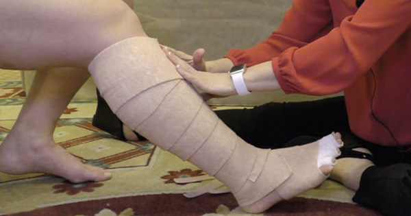
Tapes can be applied to decrease oedema and direct fluid from areas where it is not draining, towards the nearest lymph nodes or areas which are draining, by beginning applications at the desired drainage end point. Lower limb taping begins at the popliteal nodes, behind the knee, and is laid down the calf and onto the dorsum of the foot (Sobiech et al, 2022), ending near the toes, or on the toes, if cramping pains extend to the toes. A band of tape below the toes finishes the application, to stop the tapes rolling off when socks, compression or footwear are worn.
Where symptoms are chronic, to enhance lymphatic flow of the whole limb, KT can be applied from inguinal lymph nodes and laid distally down the thigh to below the knee, even if only the calf appears to require treatment. Often, as with compression, if application stops at the knee, excess fluid is drawn from the lower leg and causes oedema to collect above the knee and restrict movement (Rabe et al, 2020).
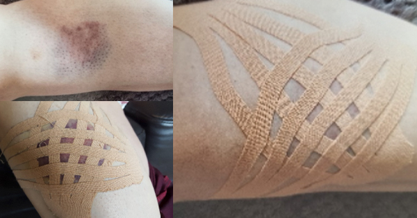
To enhance the drawing effect, fans can be laid over each other to form lattices, and further reduce tissue pressure in areas where tapes are crossed. This can focus drainage intensity by providing several different pressures within one treatment area. Stubborn oedema, bruising, haematomas and seromas are treated this way, as demonstrated in
Figure 2 (Anvar, 2015).
Even if ulcers are present, taping the periwound area will improve oxygenation of the tissues and enhance healing time. During the crucial 3-month time period, where 80% of ulcers reoccur (Probst et al, 2023), LKT can be applied onto new, fragile skin to promote healthy blood supply and prevent further necrosis caused by lack of oxygen. LKT is also useful to control pain, oedema and fibrosis in patients with post-thrombotic syndrome
(Naci et al, 2020).
Muscular movement of the limb dynamically changes the tapes’ action on the skin, alternating tissue pressures and stimulating the mechanoreceptors below. To maximise this potential, the tape is applied with the area to be taped anatomically stretched if possible:
- knee bent/ankle plantarflexed to tape on the anterior calf
- toes curled/ankle plantarflexed/ knee bent to lengthen dorsum of foot
- knee straight /ankle dorsiflexed (toes up) for posterior calf
- foot inverted to tape lateral malleolus area
- foot everted for medial foot applications.
Tape is then applied without stretch, so when the skin assumes its neutral position, tapes recoil to form corrugations which move with the skin (Sijmonsma, 2010). Consequently, with movement, a pump or micro-massage effect is created (Hamza et al, 2020). The corrugations not only deform the fascia under the tape, but influence the tension and consistency of deeper, more distant fascia, and the limb as a whole (Pamuk and Yucesoy, 2015).
Fear of doing harm
Fear of doing harm is reported as a barrier to treating high-risk patients (Elwell & Sneddon, 2019). Figures 3a and 3b demonstrate the consequence of failing to tape over a ‘liposuction burn’ caused by aggressive cannula use. The resulting necrotic wound required months of recovery and a skin graft to heal. These injuries (see Case 2) occur regularly with high volume, non-cosmetic liposuction, exacerbated by stasis caused by ill-fitting post-surgical compression garments, bunching at joints and tourniqueting blood supply to wounded tissues (Ormseth et al, 2023). Many lipoedema patients have high Body Mass Indexes (BMI), PAD, VI and comorbidities, which make them slow to heal, including diabetes and smoking (Orhurhu et al, 2021). Since this case, LKT has been applied directly over ecchymosis, regardless of cause, size or colour depth, and left in place, until desquamation removes it. Tissue breakdown is always avoided (Anvar, 2022). The following case studies document beneficial results, where ‘doing harm’ could constitute inaction.
Conclusion
Banergee (2019) reports that the main barriers to widespread use of KT are a lack of awareness or research evidence on efficacy. The paucity of literature should not preclude its use. LKT is a powerful tool, which can be applied to all symptomatic manifestations of PAD or CVI at every stage of degeneration, to reoxygenate tissues, alleviate lymphatic stasis and improve outcomes, while interacting with and addressing the venous, lymphatic and myofascial causes.
KT has few risks and treatment can be commenced even when patients are frail or ABPI contraindicates compression. LKT could halt the cycle of recurrent leg ulcers as it addresses the lymphascial involvement, which has been unreported to date.