Venous disease is common and most healthcare professionals will encounter patients who need or are receiving compression therapy, either with hosiery or bandages. Compression is the mainstay of treatment or prevention for venous ulcers and the aim must always be to ensure safe application and effective therapy. This how togu ide is intended to help practitioners understand the rationale for applying compression therapy and aid them in managing patients with lower limb venous ulceration.
Management of venous leg ulcers
Chronic venous insufficiency affects up to 50% of the adult population (Venous Forum, 2011) and it is estimated that 1% of the UK population will suffer from leg ulceration during their lifetime (Callam, 1992). The majority of these ulcers are caused by vascular disease (venous, arterial and lymphatic), with venous disease accounting for between 60-80% of leg ulceration (Callam, 1992).
Correctly applied compression therapy is now recognised as the mainstay of treatment for both the preventative and therapeutic care of venous disease, with high compression bandaging now established as the treatment of choice for venous leg ulceration (O’Meara et al, 2009). Studies would suggest that healing rates above 50% after 12 weeks of care should be achievable (Vowden et al, 1997; Barwell et al, 2004) and guidelines indicate that patients failing to respond to care within this time frame should be referred for vascular and specialist wound care assessment (Marston and Vowden, 2003).
Understanding venous disease
In a healthy individual, venous pressure at the ankle falls during exercise due to the action of the calf muscles and the presence of functional venous valves that prevent venous reflux. Figure 1 illustrates normal venous physiology and how valvular disease affects venous return. Calf muscle pump failure due to inactivity or paralysis, failure of the venous valves due to varicose veins, or damage to the deep veins secondary to venous thrombosis, trauma or venous obstruction, decrease the efficiency of this system. This results in a higher venous pressure, a condition known as chronic venous hypertension. Chronic venous hypertension has a number of consequences and changes occur in the microcirculation. This is associated with lower limb discomfort, swelling and skin changes and ultimately may lead to venous ulceration. The progression of venous disease is classified using the CEAP grading system (Eklof et al, 2004) (see Table 1).
In a normal limb a balance exists between the pressure in the capillary bed, tissue pressure and oncotic pressure. As venous pressure rises this balance is disturbed. Blood moves more slowly through the capillaries and veins increasing the risk of thrombosis and activation of white blood cells. The raised pressure also leads to increased vessel permeability with the leaking of fluid and protein into the tissues (Partsch, 2003).
Treatment of venous disease is aimed at correcting, as far as possible, the effects of valvular incompetence and reducing the damaging effects of venous hypertension. This is achieved by applying compression therapy in the form of hosiery or bandages and undertaking corrective venous surgery, endo-venous ablation therapy or sclerotherapy where investigations have demonstrated superficial venous reflux (varicose veins) to be present (Venous Forum, 2011). Treating venous disease has been demonstrated to:
- Significantly improve a patient’s quality of life
- Relieve lower limb symptoms
- Delay or prevent the long-term complications of chronic venous insufficiency
- Be a cost-effective use of resources.
- Understanding compression
- Compression therapy aims to reverse the effects of venous hypertension by:
- Decreasing the capacity of and pressure within the superficial veins. This aids venous return by increasing the blood flow velocity in the deep veins
- Reducing oedema by decreasing the pressure difference between capillaries and the surrounding tissue and transferring tissue fluid back into the vascular space. This can reduce exudate
- Minimising or reversing skin changes, to aid the healing of venous ulceration.
For compression to work it must be graduated, generating a pressure that is highest at the ankle. This must be sufficient to overcome the pressure in the lower limb veins when the patient is standing and sustained in order to deliver the necessary benefits over time. In addition, it must be tolerable to the patient.
Elastic and inelastic bandage systems
The level of compression produced by any bandage system is established by a series of complex interactions, including the size, shape and the physical structure of the limb, the type of bandage used, the layers incorporated in the bandage system, the overlap of the bandage, and the skill and technique of the bandager (Clark, 2003). Compression may contain elastic or inelastic materials or a combination of both:
- Elastic (also referred to as long-stretch) contains elastomeric fibres that are capable of stretching and returning almost to their original size.
- Inelastic (also referred to as short-stretch) contains few or no elastomeric fibres and have minimal extensibility.
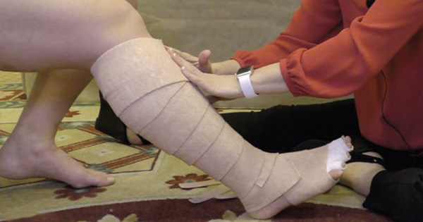
Bandages are classified according to their ability to apply and maintain a safe, predetermined level of compression (Hopkins and Worboys, 2005). When applying a bandage system to a limb the aim is to provide a ‘stiff’, but shaped container against which muscles in the calf can contract. This generates a high ‘working’ pressure, which in a graduated system, aids venous return to the heart while maintaining a lower ‘resting’ pressure during inactivity (Fig 2).
Stiff bandaging may be achieved by using either inelastic bandages or elastic bandages in a multi-layer system such as the 4-layer bandage (Partsch, 2005). Elastic bandages are rarely used in isolation in current practice, as they provide little or no stiffness. For a table showing the classifications for the different compression bandages see Beldon, 2009.
One of the beneficial effects of compression with a high static stiffness index (SSI) (Fig 3) is a rapid initial change in limb size as oedema is reduced. One consequence of this is that the bandage system will need to be re-applied more frequently due to the rapid reduction of oedema. This is particularly relevant when using inelastic materials as, without elastomeric fibres, their effectiveness will decrease and bandage slippage is more likely to occur as these systems fail to accommodate for the change in limb circumference. The importance of SSI in restoring venous function is shown in Figure 4.
Understanding compression levels
Bandage systems and compression hosiery are graded according to the level of compression they generate. Several classification systems exist. To avoid confusion when describing the level of compression applied to the limb, WUWHS suggests using the following terminology (2008):
- Mild (less than 20mmHg)
- Moderate (20-40mmHg)
- Strong (40-60mmHg)
- Very strong (greater than 60mmHg).
Strong compression (>40mmHg) is generally recommended for the treatment of a venous leg ulcer. For some patients factors such as mild arterial disease, neuropathy or cardiac failure render strong compression unsafe or painful and mild or moderate compression may be required (eg using inelastic compression). Patients with more severe arterial disease should not receive compression (Marston and Vowden, 2003).
Factors affecting sub-compression system pressure
Using a compression system (bandages or hosiery) alone does not guarantee a level of compression. Pressure will vary according to the limb size and shape, level of calf muscle activity, the bandage or hosiery characteristics, bandage width and the degree of overlap and the application tension. Many of these factors are controlled by the bandager whose skills will also affect the graduation of compression and the comfort and durability of the bandaging.
Most bandage systems give advice on variations to accommodate differing ankle circumference. Larger limbs, ie those with a higher circumference, will require a higher classification bandage to exert the desired level of compression. Similar constraints apply to hosiery. Careful measurement and fitting is important as is the choice of hosiery itself; the characteristics of circular (elastic) and flat knit (inelastic) differ in a similar way to bandages.
Assessing patients before application
Arterial assessment using Doppler ultrasound to calculate the ankle brachial pressure index (ABPI) should be undertaken before considering compression therapy (SIGN, 2010; Vowden and Vowden, 2001). The ABPI defines both the level of compression and the need for onward referral to vascular specialists. In addition, before selecting patients for application of compression, assess the skin condition and limb shape, as well as the presence of neuropathy or cardiac failure and patient-known allergies as these may affect both the level of compression and the components of the compression system used (Marston and Vowden, 2003). The assessment process should identify potential problems that may affect healing and recurrence.
Assessment should also identify potentially vulnerable areas such as bony prominences, which may require padding for protection. Padding may add to the comfort of the bandage but excessive padding may reduce compression levels and lead to bandage slippage and generate a bulky bandage system. When assessing for hosiery, consider the patients’ and their carers’ ability to apply and remove stockings.
Choosing and applying compression
A recent Cochrane review has identified that multi-component
bandage systems have been shown to be more effective than single-component bandage systems (ranging from cohesive single layer through to zinc paste bandages and Unna’s boot) in healing venous leg ulcers (O’Meara, 2009). Box 1 highlights the requirements for an ideal compression system.
The choice of compression system for each patient will depend on the results of the assessment process, the patient’s preferences, healthcare professionals’ skills and the available resources. Effective compression should provide a balance between exerting too little pressure, which is ineffective, and too much pressure, which causes damage or is not tolerated by the wearer. Other considerations may relate to the bulk of the bandage, the impact on footwear and temperature discomfort during hot weather. If the chosen bandage system is bulky ensure that the patient has or is provided with suitable footwear. This will encourage mobilisation, increase the effectiveness of treatment and aid concordance.
For compression to be fully effective, patients should be provided with appropriate education on the underlying disease and be encouraged to elevate their legs when resting (WUWHS, 2008).
Choosing and using hosiery
Hosiery remains the mainstay of prevention, but 2-layer ‘strong’ hosiery systems can also be an effective method of providing compression therapy in selected patients with small, low exudate wounds. Such patients can be encouraged to self-care under the supervision of an appropriate healthcare professional. Provision of application aids can help to facilitate this.
It is important to measure the leg accurately and select appropriately sized hosiery with the correct compression level. Ensure that the patient is shown how to apply the hosiery and understands when to wear the garment. Give instruction on skin care and hosiery maintenance, including washing and drying. Hosiery constituents vary and it may be necessary to try different makes of stocking if concordance is to be improved for individual patients.
Improving concordance
A patient’s initial experience with compression therapy may colour their subsequent opinion of this form of therapy. Patients should be engaged in treatment planning and be provided with sufficient information for them to understand the rationale for treatment. Adherence with treatment is also dependent upon patient motivation, which can be affected by factors such as social isolation or treatment discomfort. Effective symptom control, either with dressings or analgesia, can improve quality of life and patient tolerance of compression therapy, aiding concordance (Briggs and Nelson, 2010). Miller et al (2011) in their study on predicting concordance concluded that pain, wound size and depth and patient age all influenced concordance.
For further guidance on the use of compression therapy in venous leg ulcers, please see the recommended treatment pathway developed by the International Leg Ulcer Advisory Board (Marston and Vowden, 2003. Available from www.woundsinternational.com)
References
- Barwell JR, Davies CE, Deacon J, et al (2004) Comparison of surgery and compression with compression alone in chronic venous ulceration (ESCHAR study): randomised controlled trial. Lancet 5;363(9424):1854-9
- Beldon P (2006) Bandaging: which bandage to use and when. Wound Essentials 2006;4:52-61
- Briggs M, Nelson EA (2010) Topical agents or dressings for pain in venous leg ulcers. Cochrane Database of Systematic Reviews 4:CD001177
- Callam M (1992) Prevalence of chronic leg ulceration and severe chronic venous disease in western countries. Phlebology 7(Suppl):6-12
- Clark M (2003) Compression bandages: principles and definitions. In: EWMA Position Document. Understanding compression therapy. London: MEP Ltd. 5-7
- Eklof B, Rutherford RB, Bergan JJ, et al (2004) Revision of the CEAP classification for chronic venous disorders: consensus statement. J Vasc Surg 40(6):1248-52
- Hopkins A, Worboys F (2005) Understanding compression therapy to achieve tolerance. Wounds UK 1(3):26-34
- Marston W, Vowden K (2003) Compression therapy: a guide to safe practice. In: EWMA Position Document. Understanding Compression Therapy. London: MEP Ltd. 11-7
- Miller C, Kapp S, Newall N, et al (2011) Predicting concordance with multilayer compression bandaging. J Wound Care 20(3):101-2,4,6 Passim
- Moffatt CJ (2004) Factors that affect concordance with compression therapy. J Wound Care 13(7):291-4
- O’Meara S, Cullum NA, Nelson EA (2009) Compression for venous leg ulcers. Cochrane Database of Systematic Reviews 1:CD000265
- Partsch H (2003) Understanding the pathophysiological effects of compression. In: EWMA Position Document: Understanding compression therapy. London: MEP Ltd. 2-4
- Partsch H (2005) The use of pressure change on standing as a surrogate measure of the stiffness of a compression bandage. Eur J Vasc Endovasc Surg 30(4):415-21
- SIGN (2010) Management of Chronic Venous Leg Ulcers. Edinburgh: Scottish Intercollegiate Guidelines Network
- Venous Forum of the Royal Society of Medicine. Berridge D, Bradbury AW, Davies AH, et al (2011) Recommendations for the referral and treatment of patients with lower limb chronic venous insufficiency (including varicose veins). Phlebology 26(3):9-3
- Vowden KR, Barker A, Vowden P (1997) Leg ulcer management in a nurse-led, hospital-based clinic. J Wound Care 6(5):233-6
- Vowden P, Vowden K (2001) Doppler assessment and ABPI: Interpretation in the management of leg ulceration. World Wide Wounds, March.
- World Union of Wound Healing Societies (WUWHS) (2008) Principles of best practice: Compression in venous leg ulcers. A consensus document. London: MEP Ltd.
Authors: Vowden K, Nurse Consultant Wound Care; Peter Vowden, Vascular Surgeon and Visiting Professor of Wound Healing Research. Bradford Teaching Hospitals NHS Foundation Trust and The University of Bradford

