
Lower limb ulceration is commonly managed within community settings, where nurses and tissue viability teams are often the first clinicians to identify underlying arterial disease (Pagnamenta et al, 2024). However, arterial insufficiency remains frequently under-recognised or misinterpreted, leading to delayed healing, inappropriate compression therapy and avoidable patient harm (All-Party Parliamentary Group on Vascular and Venous Disease, 2019). Due to the increased fear of harm through inappropriate care, the need to exclude peripheral arterial disease (PAD) is, therefore, fundamental to safe lower-limb ulcer management.
Arterial duplex scanning provides detailed information on arterial anatomy and haemodynamics (Zubair and Lotfollahzadeh, 2023). However, many clinicians working in community settings report uncertainty in how duplex findings should be interpreted and translated into clinical decision-making. As access to duplex imaging becomes more widespread, it is essential that clinicians develop confidence in understanding the information provided within these reports. This article provides a practical guide to understanding arterial duplex ultrasound reports in the context of lower limb ulcer assessment. It explains the principles of duplex imaging, describes common report terminology, and highlights how duplex findings should be interpreted alongside bedside vascular assessment to inform safe clinical decision-making.
Duplex scanning role within community nursing
Arterial duplex scanning can be an important investigation within lower-limb care pathways, but it does not replace a thorough individualised patient assessment. Clinical examination remains the foundation of vascular assessment and includes palpation of pedal pulses, assessment of handheld Doppler sounds/waveforms, measurement of ankle–brachial pressure index (ABPI) or toe brachial pressure index (TBPI), and consideration of the patient’s medical history and symptoms (NICE, 2018). These assessments help identify patients who may have underlying arterial disease and determine when further vascular imaging may be required.
In community practice, duplex imaging is usually requested when initial clinical findings raise concern for arterial insufficiency. However, awareness of duplex as a diagnostic tool remains variable among community clinicians.
Duplex imaging may be appropriate when:
- ABPI or TBPI values suggest arterial disease.
- Doppler waveforms are monophasic or difficult to interpret.
- A lower limb ulcer is unusually painful or atypical in appearance.
- Ulcers fail to progress despite appropriate compression therapy.
- Symptoms of peripheral arterial disease are present (e.g. intermittent claudication or rest pain; College and Society for Clinical Vascular Science, 2021).
Duplex ultrasound provides detailed imaging of arterial structure and blood flow that complements holistic patient assessment. It effectively provides a map of arterial blood flow, allowing clinicians to understand not only whether blood is reaching the limb but also how effectively it is doing so. In simple terms, a handheld Doppler allows clinicians to listen to blood flow, whereas duplex imaging allows clinicians to visualise what is happening within the artery itself. A normal duplex result provides reassurance that arterial perfusion is adequate and that best-practice compression therapy can usually be applied safely. Conversely, abnormal findings should prompt clinicians to review compression therapy, escalate concerns and consider referral for vascular specialist assessment to ensure safe and appropriate patient management.
Arterial duplex scanning
When PAD is suspected, duplex ultrasonography can be used to map the arterial tree and support vascular treatment decision-making (Hodgkiss-Harlow and Bandyk, 2013). A normal duplex scan confirms adequate arterial perfusion and supports clinical decision-making regarding compression therapy. Duplex ultrasonography is a non-invasive imaging modality with high diagnostic accuracy, demonstrating approximately 80% sensitivity and 90–100% specificity for detecting large-vessel arterial disease (Donnelly et al, 2000).
By combining real-time ultrasound imaging with Doppler waveform analysis, arterial duplex scanning provides information on both arterial structure and blood flow. It allows clinicians to visualise the condition of the arterial wall and assess how effectively blood is moving through the vessel lumen (Zubair and Lotfollahzadeh, 2023). Any artery located sufficiently close to the skin surface can be examined using duplex ultrasound, making it particularly valuable in lower limb vascular assessment. In simple terms, duplex imaging enables clinicians to determine the location, extent and severity of PAD. A lower limb arterial duplex examination typically begins at the common femoral artery and proceeds distally through the profunda femoris artery, superficial femoral artery, popliteal artery and the tibial and pedal arteries [Figure 1]. Where clinically indicated, the examination may also extend proximally to include the aorta and the common and external iliac arteries (Society for Vascular Technology of Great Britain and Ireland, 2022).
Arterial duplex ultrasonography consists of three key components: B-mode imaging, colour Doppler and spectral Doppler. B-mode imaging (also referred to as greyscale imaging) uses high-frequency sound waves to produce real-time anatomical images of the artery. On B-mode imaging, the vessel typically appears as a dark tubular structure, while atherosclerotic plaque or areas of stenosis appear brighter (white or grey) due to increased echogenicity (Zubair and Lotfollahzadeh, 2023). B-mode assessment enables visualisation of arterial wall anatomy, luminal patency, plaque presence and morphology, and vessel diameter (Zwiebel and Pellerito, 2005).
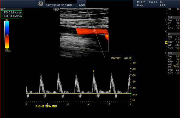
Colour Doppler imaging provides additional information on the direction and relative velocity of blood flow within the artery. This helps identify areas of disturbed or altered flow that may be associated with arterial narrowing or occlusion (El-Sayed et al, 2020). Spectral Doppler complements this by displaying blood flow velocity over time as a waveform. Higher peak velocities indicate faster blood within the artery; this waveform is typically characteristic of normal arterial flow [Figure 2]. In contrast, spectral broadening (filling in of the window below the waveform) reflects disturbed or turbulent flow, which may indicate haemodynamically significant stenosis (Nasra and Osher, 2025).
The integration of B-mode, colour Doppler and spectral Doppler imaging [Figure 2] enables clinicians to assess both the anatomical structure of the artery and the haemodynamic characteristics of blood flow. This combined approach improves the diagnostic reliability of duplex ultrasonography for detecting arterial stenosis or occlusion. A summary of these components is provided in Table 1.
Limitations of arterial duplex scanning
Arterial duplex ultrasonography is a valuable investigation in the assessment of lower-limb arterial disease; however, its accuracy may be influenced by several technical and patient-related factors. These variables can affect image quality and, in some cases, limit the completeness or reliability of the assessment. The following limitations are commonly encountered in clinical practice:
- Patients with a high BMI: A high BMI can make duplex scanning more challenging and time-consuming. Increased subcutaneous adipose tissue increases the distance between the probe and the artery, weakening returning ultrasound signals and reducing image resolution (Nabasenja et al, 2022).
- Presence of wounds, dressings, bandages or casts: Duplex scanning requires direct probe-to-skin contact. Dressings, compression bandaging or casts can obstruct access to the limb, and many vascular laboratories do not have the ability to safely remove and reapply these products. If these cannot be removed prior to the appointment, the examination may be significantly limited (Society for Vascular Technology of Great Britain and Ireland, 2022).
- Recent surgical intervention: Postoperative oedema, haematoma, tenderness or surgical staples may restrict transducer placement and temporarily reduce image quality, limiting the ability to obtain a complete assessment.
- Calcified plaque: Heavily calcified atherosclerotic plaque, commonly seen in patients with diabetes or chronic kidney disease, produces acoustic shadowing that obscures the arterial lumen and limits accurate Doppler velocity measurement (Mohebali et al, 2015).
- Patient positioning and cooperation: Successful duplex imaging requires patients to maintain specific limb positions. Pain, reduced mobility, breathlessness, joint contractures, neurological disease, or cognitive impairment may impair cooperation, resulting in incomplete visualisation or reduced image quality.
- Difficulty assessing distal and pedal vessels: Distal and pedal arteries are critical when assessing perfusion in patients with ulceration, but are often small, calcified, and obscured by oedema, tissue loss or dressings (Hartshorne, 2001).
It is important to recognise that arterial duplex ultrasonography assesses large-vessel patency and haemodynamics but does not evaluate microvascular perfusion. Consequently, microvascular impairment may still be present in patients with diabetes or chronic limb-threatening ischaemia despite normal proximal arterial findings (Conte et al, 2019). Arterial duplex findings should therefore always be interpreted within a holistic assessment framework, alongside clinical examination, ankle brachial or toe brachial pressure measurements, and WIFI classification to ensure accurate evaluation of patients with suspected arterial disease (Hinchliffe et al, 2020).
Arterial duplex report interpretation
Interpreting a lower limb arterial duplex report should always be considered in the context of the patient’s presenting symptoms and clinical findings. Providing clear clinical information on the imaging request is essential, as this guides the vascular sonographer in targeting areas of concern and determining the scope of the examination.
The duplex report will include an assessment of arterial waveforms, also referred to as spectral waveform analysis, for each vascular segment examined. Doppler waveform analysis is essential for understanding both the presence and severity of arterial disease (Kim et al, 2020). Although duplex scanning allows more detailed evaluation of waveform morphology, classification remains consistent with that used during handheld Doppler assessment for ABPI measurement. Waveforms are typically categorised as:
- Triphasic – characterised by a sharp systolic upstroke, early diastolic flow reversal, and late diastolic forward flow.
- Biphasic – loss of early diastolic flow reversal, indicating early haemodynamic change.
- Monophasic – reduced systolic flow with loss of both early diastolic reversal and late diastolic forward flow, suggesting significant arterial disease.
In clinical practice, monophasic waveforms typically indicate haemodynamically significant arterial disease and should prompt careful review of compression therapy and consideration of vascular referral.
The duplex report will also describe the anatomical level and severity of disease within each arterial segment. Disease severity is quantified by the degree of luminal narrowing, referred to as diameter reduction (DR) [Table 2]. Stenoses greater than 50% are generally considered haemodynamically significant and may affect distal perfusion, particularly when located within major inflow vessels such as the iliac or femoropopliteal segments.
At this stage of the examination, the vascular sonographer identifies the presence and anatomical level of arterial disease by assessing areas of reduced blood flow within the arterial lumen. Focus is then directed to segments where stenosis or occlusion is suspected, and peak systolic velocity (PSV) measurements are obtained. PSV, measured using spectral Doppler ultrasound, represents the maximum blood flow velocity during the systolic phase of the cardiac cycle and is expressed in centimetres per second (cm/s). Assessment of flow velocity enables detection and quantification of arterial narrowing, as velocities typically increase at sites of stenosis due to luminal constriction.
To further assess and quantify the degree of stenosis, the peak systolic velocity ratio (PSV ratio) is calculated. This is determined by dividing the PSV measured at the stenotic lesion by the PSV recorded in a normal arterial segment proximal to the stenosis. The PSV ratio provides an objective measure of haemodynamic significance and is used to grade the severity of arterial narrowing [Table 3]. By combining measurements of vessel diameter and blood flow velocity, duplex scanning provides insight into both the location and haemodynamic severity of arterial disease.
Duplex reports may also include comments on collateral flow, which occurs when an arterial segment becomes significantly narrowed or occluded, and blood is redirected through smaller alternative vessels. Collateral circulation [Figure 3] develops when blood is redirected through smaller vessels to bypass an obstructed artery. These vessels act as natural bypass pathways and may help maintain distal perfusion in chronic arterial disease. The presence of well-developed collateral vessels often indicates long-standing disease, whereas acute occlusion is usually associated with minimal collateralisation (McDermott et al, 2014).
The vascular sonographers will conclude the report by summarising key findings and highlighting clinically significant disease. This typically includes identification of the affected arterial segments, description of disease severity, and supporting haemodynamic data such as measured velocities or PSV ratios. Examples of report terminology include:
- Calcified plaque at the origin and distal common iliac artery (CIA) bifurcation without significantly elevated velocities.
- Common femoral artery patent with mild–moderate disease and preserved triphasic flow.
- Superficial femoral artery patent but diseased, with increasing velocities through the mid-to-distal thigh (PSV 122–388 cm/s)
- Popliteal artery patent above the knee with a 3.2 cm occlusion and distal reconstitution; weak distal flow (PSV 11 cm/s).
The duplex report enables clinicians to correlate imaging findings with clinical examination. While clinical assessment reflects the functional impact of disease on the patient, duplex imaging provides detailed anatomical and haemodynamic information, allowing comprehensive evaluation and informed clinical decision-making.
Conclusion
Arterial duplex ultrasound provides valuable anatomical and haemodynamic insight into lower limb arterial disease. However, the value of this investigation depends on clinicians’ ability to interpret the findings within the context of holistic vascular assessment.
Community nurses and tissue viability teams are frequently the first clinicians to recognise arterial insufficiency in patients with lower limb ulceration. Strengthening vascular literacy within the community workforce will support safer compression therapy, earlier identification of peripheral arterial disease and timelier referral to vascular services. Ultimately, improving clinicians’ confidence in interpreting duplex reports has the potential to significantly improve healing outcomes and patient safety.